2D et 3D imaging, live samples, images analysis
The C3M imaging facility provide helps , training, technological and biological developpement for cell and tissue imaging to scientists comming from the plubilc or private companies.

Microscopy network Imagerie Côte d’Azur
The C3M microscopy platform is labeled Infrastructure in Biology, Health and Agronomy (IBiSA). It is a member of the Microscopy Imaging Côte d'Azur (MICA) network which pools the technical platforms (light and electronic microscopy, cytometry, image analysis) of 8 partners in academic research on the Côte d'Azur.


Imaging manager Marie Irondelle
I train you on the devices of the platform.
I can help you develop protocols.
I answer your questions about the operation or the technical possibilities of the platform.
marie.irondelle@univ-cotedazur.fr

Imaging Engineer Anne Doye
I can help you find technical solutions and train you on the platform's devices.
anne.doye@univ-cotedazur.fr
To access the platform, users must have been trained by platform staff and sign the user charter and one training sheet per device. Users must register for each device on the C3M intranet. The platform can provide specific imaging media (multi-well ibidi, fluorodish, etc.) or antibodies needed when implementing new protocols.
As stipulated in the charter, publications containing data generated on the platform must mention it in the acknowledgments.

This microscope is equipped with a Nikon DS-L3 color camera and is dedicated to the visualization of histological sections stained with different contrast methods (carmine red, hematoxylin/eosin, Masson's trichrome...).
Specifications techniques
This upright widefield microscope from Leica is equipped with an ORCA-ER camera (Hamamatsu) and allows routine fluorescence imaging.
Specifications techniques
This widefield microscope from Zeiss is equipped with a Flash4 camera (Hamamatsu) and a thermostat-controlled chamber (CO2 and temperature control) for routine fluorescence imaging of living samples.

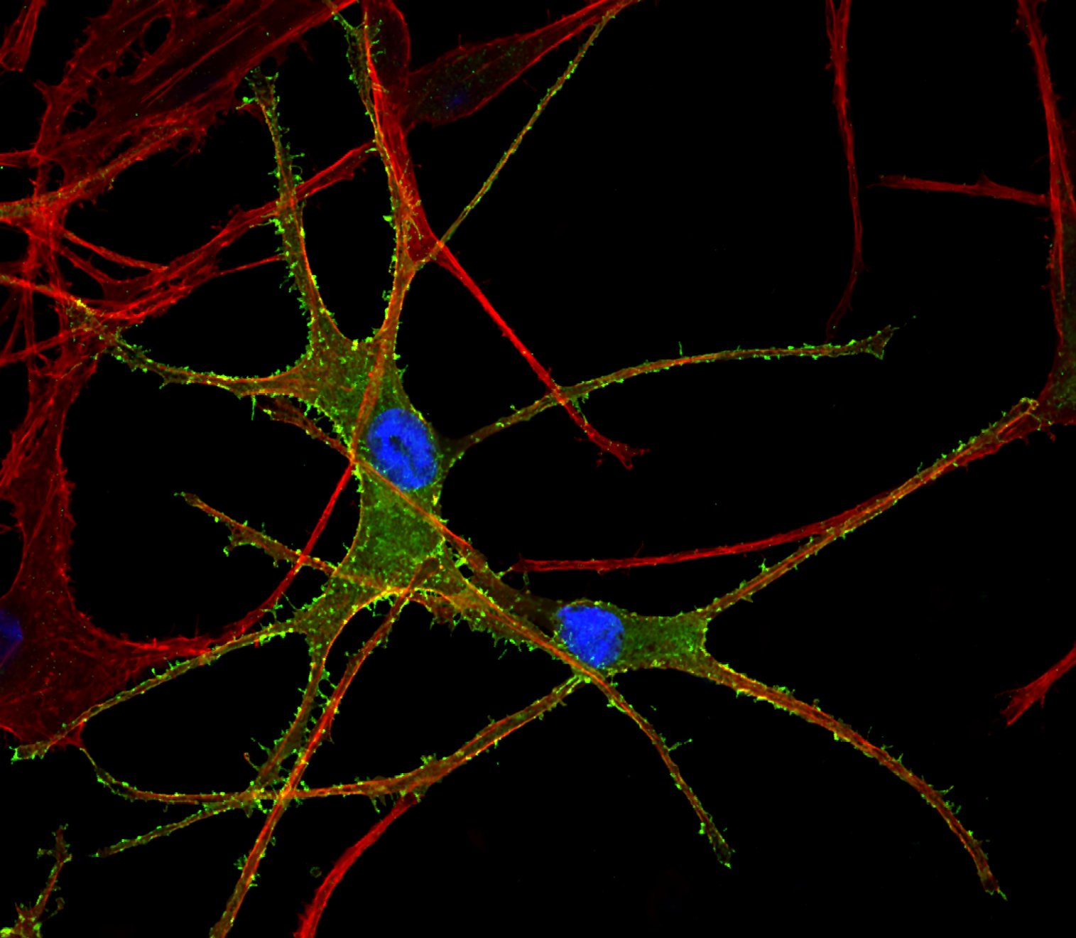
Confocal microscopy offers several advantages over conventional optical microscopy, including shallow depth of field, elimination of out-of-focus blurry, and the ability to collect serial optical sections from thick specimens.
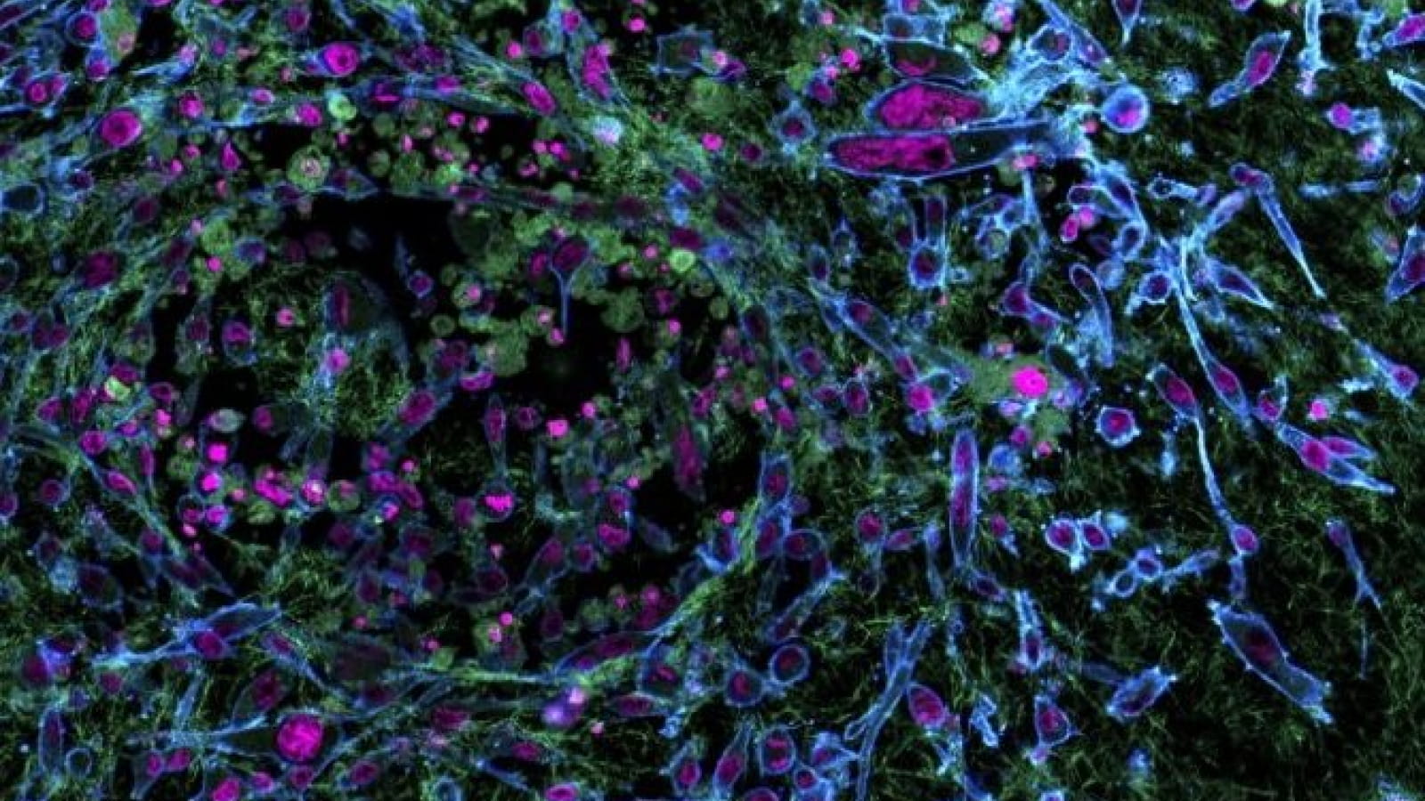
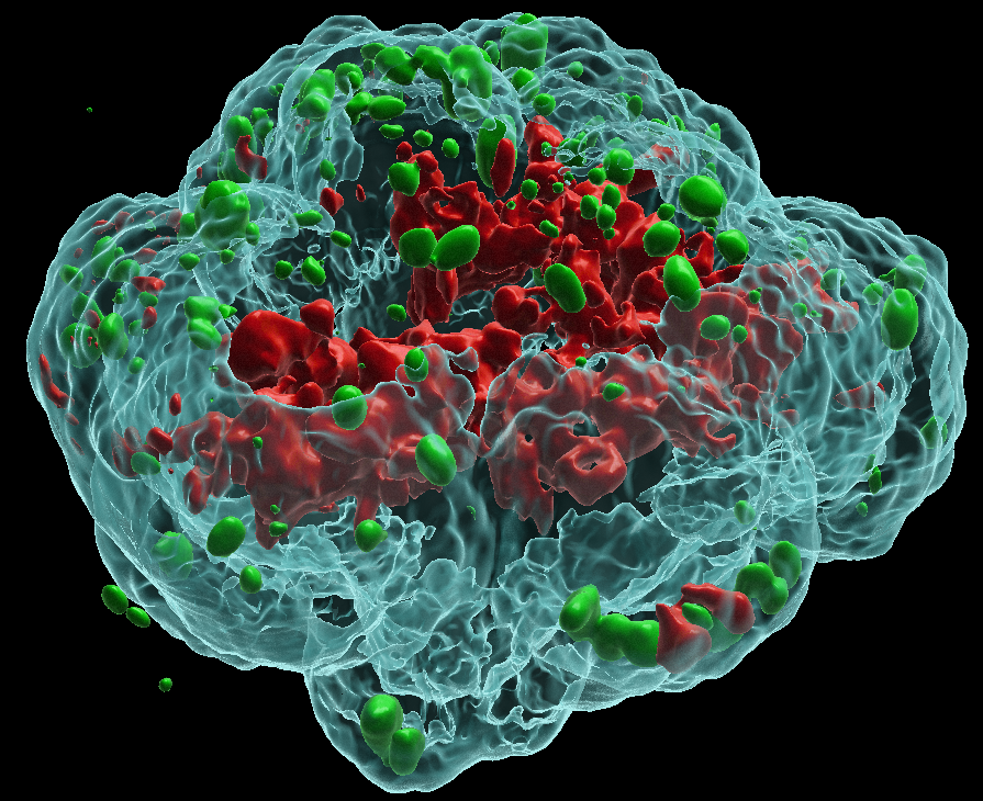
This microscope allows the acquisition of thick samples such as tissues, spheroids or organoids, as well as the acquisition of small intracellular organelles such as endosomes, the Golgi or the mitochondrial network.

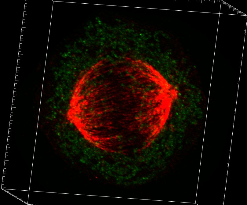
The spinning disk combines the speed of acquisition with the resolution of a confocal. This microscope enables thick samples to be acquired with the precision of a confocal microscope and the speed of a wide-field microscope. Simultaneous illumination of the entire observation zone reduces acquisition time and photo-toxicity. This system is therefore particularly well suited to the acquisition of live samples. It is equipped with a thermostat-controlled chamber (temperature and CO2 control).
Technical specifications| Inserm | Académique (hors Inserm) | Privé | |
|---|---|---|---|
| Use of equipment (per hour - 1 hour minimum) | 38,84€ | 51,73€ | 80€ |
