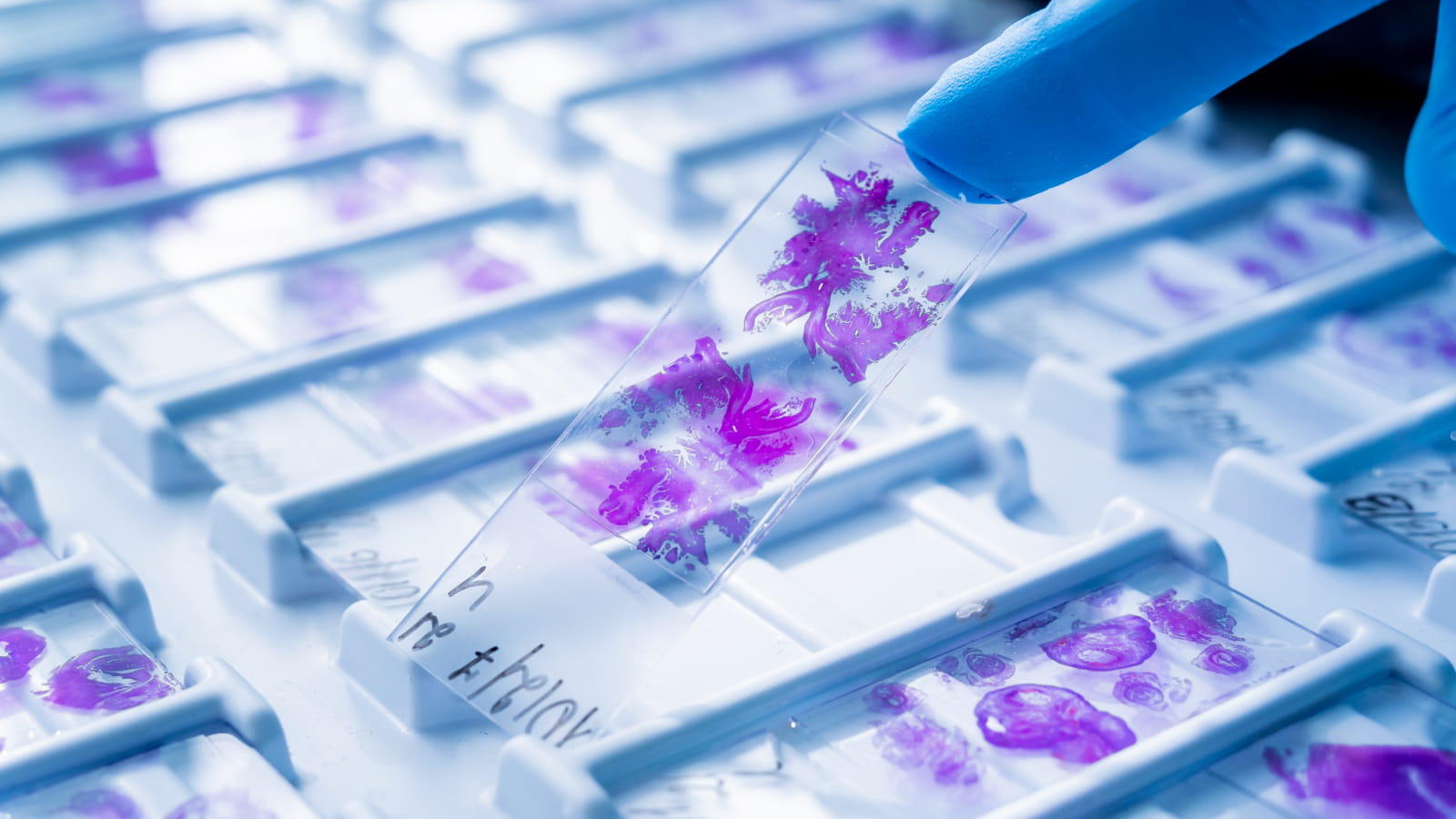
Histology, Immunolabelling and Tissue Clearing
The C3M histology facility HistoC3M was created in 2013 with funding from the Canceropôle PACA, which allowed the purchase of state-of-the-art equipment. The histology facility is equipped to embed tissues in paraffin, cut paraffin blocks or frozen tissues, clear tissues and perform automated immunostaining and histological staining. The HistoC3M platform has many functions: it ensures maintenance of the equipment, provides users with theoretical training in both standard (cryostat, paraffin, etc.) and innovative histology approaches (tissue clearing) and offers practical training in the operation of the devices, and scientific and technical advice. It also maintains a technological watch and is involved in innovation and development, etc.

Scientific Manager Jérome GILLERON
Please contact me if you have any questions about the operation
or technical possibilities of the platform.Phone : 04 89 15 38 31
Jerome.GILLERON@univ-cotedazur.fr

To access the HistoC3M facility, users must be trained by the staff and sign the user charter. Users must register for each device on the C3M intranet. The platform pools the purchase of solvents and standard stains, paraffin, disposable knives and superfrost glass slides. Everything else is paid by the user labs.
As stated in the charter, publications containing data generated using the HistoC3M facility must mention in their acknowledgements: "Histology analyses were performed at HistoC3M, a fully equipped histology facility funded by the Canceropole PACA."
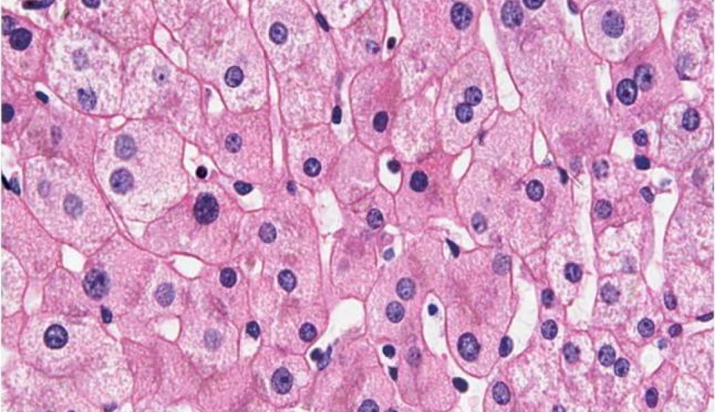
Haematoxylin and eosin staining (HE staining) is a commonly used staining technique in histology and histopathology.
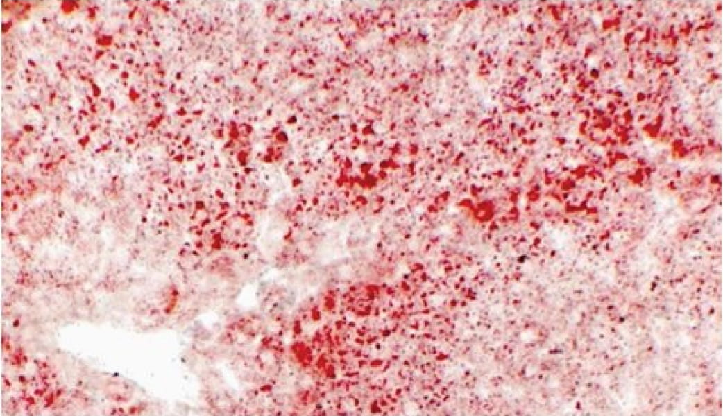
Oil Red O is a stain used to visualize neutral triglycerides and lipids.

Immunofluorescence is a labelling technique that uses antibodies coupled to fluorochromes.
The STP120 Spin Tissue Processor is an automated machine that can coat up to 100 cassettes at the same time. The fixed tissues are dehydrated by increasing baths of alcohol (70, 80, 95 and 100%). The alcohol is then replaced by xylene, a solvent with a better exchange capacity. Finally the tissues are incubated with a hot paraffin that infiltrates the tissues. The tissues are then embedded using the EC 350 Modular Tissue Embedding Center.
The Modular Tissue Embedding Center EC 350 is a hot paraffin dispenser used to embed tissues already coated with paraffin by the STP120. Tissues are extracted from the cassettes, placed in a mold which is then filled with paraffin and mounted in a block together with the bottom part of the cassette. The blocks are then cooled on a cold plate included in the system.
The Microtome HM340E is equipped with the Niagara system which allows the paraffin sections to be directly collected in a hot water bath. This system allows users to obtain sections that faithfully preserve the structure of the tissues but it also makes it simpler for occasional users to cut sections. This microtome cuts paraffin-embedded tissue sections of about 1 to 25 µm thickness that can be used for histological staining and/or immunostaining analyses (although frozen sections are more suitable for immunostaining).
The PT antigen retrieval module is used to heat solutions in which samples are immersed in order to retrieve some of the antigenicity of paraffin tissue sections. The solutions used are citrate buffer pH6 and Tris-EDTA pH9.
The Labvision 350 Auto-Immunostainer automates the labelling of paraffin and/or frozen tissue sections in a simple, efficient and reproducible way.
The Myreva autostainer simultaneously and automatically performs standard histological staining on large series of slides. The standard stains used are Hematoxylin/Eosin, Oil-Red-O and Alcian Blue, but any new staining protocol can be added on request by sending an e-mail to the facility managers.

The Cryostat CM350 freezes sections of tissue embedded in OCT (or tissue Tek). The sections frozen at -14°C to -55°C (depending on the tissue) preserve the tissue antigens in an optimal way. The morphology of tissues is not as good as those embedded in paraffin sections but remains very acceptable.
| Academic | Private | |
|---|---|---|
| Device use (per hour - 1h minimum) | €77 | €100 |
| Mandatory training on each device | €50 | €100 |
| Theoretical training in histology (1h30) | €50 | €100 |

Everything you always wanted to know about histological staining and immunostaining but were afraid to ask: from sample preparation to observation.


The HistoC3M facility has developed a tissue transparency technology and a dedicated imaging and analysis pipeline. For more information, please contact the HistoC3M scientific manager.